The Flow State - Neuroanatomy Self-Portrait


Tools: Maya, Adobe Illustrator, Procreate,
Adobe Photoshop
Client: Dr. Shelley Wall
Target audience: Lay Audience
December 2025
Goal: The goal was to create the brain in-situ of a self-portrait. The brain had to be shown in an interesting cross section and could follow a path or theme. I focused on the mechanism of the flow state!
You might have experienced the flow state at some point of your life - where the task you’re focusing on feels effortless and your body and mind is completely absorbed and in focus. It generally occurs when you present yourself in enganging and challenging activities.
Ideation & Maquettes
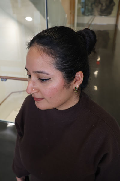
When I started first took my reference photo for the self-portrait, I wanted to angle it so the ventral tegmental area and substantia nigra are visible in my cross-section. This is because I wanted to highlight the regions which produce dopamine. I also wanted to experiment with different cross sections at multiple levels of the brain, thus I utilized 3D softwares to create maquettes.


Progress of Sketches & Feedback

By continuing to think of the story I wanted to communicate for this piece, I researched the most important regions of the flow state I wanted to highlight. I found it very interesting that the frontal lobe which is typically always active, is not as active in the flow state. Instead, the cerebellum takes over and increases in activity.
From this research, I still wanted to show a portion of the frontal lobe while still showing the substantia nigra and ventral tegmental area and the cerebellum. I accomplished this by creating multiple cross sections (horizontal, coronal and sagittal) to create an interesting view.
Feedback from peers:
-
Make basal ganglia bigger
-
Skull cross section needs to be fixed near the occipital lobe
-
Fix corpus callosum region, it is not as curved
I spent lots of time also looking at the Human Central Nervous System atlas for cross section and the UBC cross sections (horizontal and coronal to represent the accurate anatomy.


References
-
Alameda, C., Sanabria, D., & Ciria, L. F. (2022). The brain in flow: A systematic review on the neural basis of the flow state. Cortex, 154, 348–364. https://doi.org/10.1016/j.cortex.2022.06.005
-
Horizontal brain slices. Functional Neuroanatomy. University of British Columbia. https://neuroanatomy.ca/horizontals.html
-
Coronal brain slices. Functional Neuroanatomy. University of British Columbia. https://neuroanatomy.ca/coronals.html
-
Nieuwenhuys, R., Voogd, J., & Van Huijzen, C. (2019). Human Central Nervous System (4th ed.). Springer.
-
Rohen, Johannes W., et al. Color Atlas of Anatomy: A Photographic Study of the Human Body. 7th ed., Williams & Wilkins, 2011.
-
Schünke, Michael., Schulte, Erik., & Schumacher, Udo. (2010). Thieme atlas of anatomy. Head and neuroanatomy. Thieme.
-
van der Linden, D., Tops, M., & Bakker, A. B. (2021). The Neuroscience of the Flow State: Involvement of the Locus Coeruleus Norepinephrine System. Frontiers in Psychology, 12. https://doi.org/10.3389/fpsyg.2021.645498