Low-Grade Ovarian Serous Carcinoma
Low-Grade Ovarian Serous Carcinoma

Tools: Adobe Illustrator, Procreate, Autodesk Maya
Client: Prof. Dave Mazierski & Dr. John Wang
Target audience: Educated lay audience
April 2026
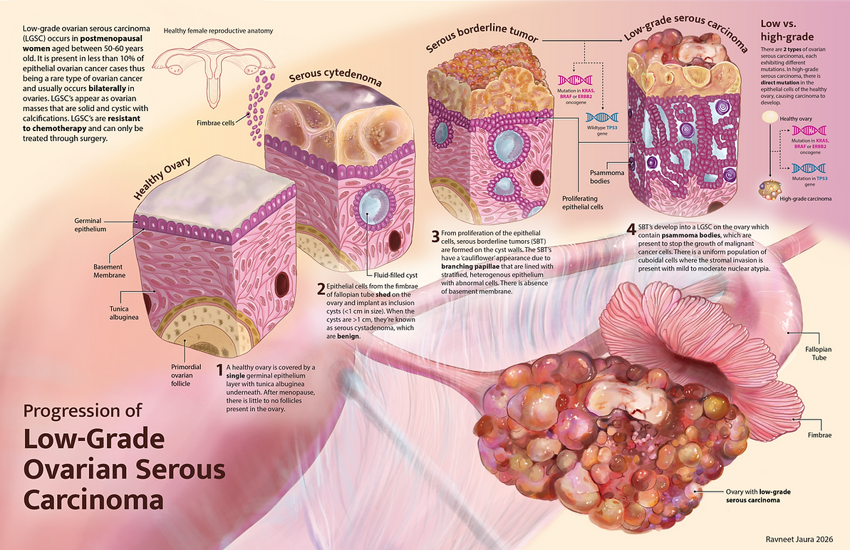
Goal: This 2-page spread is to communicate about low-grade serous ovarian carcinoma and show the progression of this cancer from a healthy ovary towards the low-grade cancer. The progression of the changes in the cellular scale is shown through tissue cubes to communicate clearly which specific changes are occurring. The final stage of how the ovary appears at the cancerous stage largely takes over the 2nd page to capture the viewers attention. Finally, the difference between low-grade and high-grade was important to highlight in terms of the mutation differences.
Research
The first step of this project was to extensively research in the chosen topic. I selected ovarian cancer, due to my interest in female reproductive biology desire to understand the anatomy and underlying science. Through my research, I learned there were two primary types of ovarian cancers - serous and mucinous and I explored the key differences between them. When I first approach a project, I make sure all the research is accurate and I ensure I have a strong knowledge base before moving into my visual work.

Sketches & Iteration

As I had researched, the progression to the low-grade ovarian cancer was through 4 steps. Representing the details of each step was done best through tissue cubes. The main differences in the pathology also occurred on the surface of the ovary, which was important to highlight. I chose an angle where the interior of the cubes and the exterior surface could be shown.
Once the tissue cubes were approved, I moved on to the overall layout and visual style of the piece. I knew I wanted to include a full ovary illustration, and the most appropriate choice was one that depicted the ovary alongside the pathology being communicated. Since there is a sequence to the pathology of the story, I added letters from A-D to clarify reading order - this was later changed to numbers 1-4.
Layout Version 1:

In the first layout, the focal point began on the left, with the tissue cubes read from bottom to top. The primary issue with this layout was the reading order, and the angle of the cubes was not the most visually compelling.
Layout Version 2:

In the second layout, I spaced the tissue cubes across both pages to establish a clearer reading order, and aligned the final cube with the ovary pathology for a more cohesive composition. However, the spacing between cubes was uneven, and the ovary illustration spanned the full page, clashing with other important areas of interest.
Rendering & Layout
Moving into the rendering phase, I referenced multiple sources to accurately depict the pathology at each stage. The epithelial cells were the primary point of interest, so I went through several colour iterations to find a palette that was salient without overpowering the composition. At the same time, I was refining the ovary rendering and testing colours that would feel cohesive across both elements. For the uterine tissue, I chose a light pink with blue-grey shadows to convey a sense of translucency, and carried the same blue tone into the cyst fluid to maintain colour harmony throughout the piece.
Test renders:


Final renders of tissue cubes and uterus with low-grade serous carcinoma ovary:


Low-grade vs. high-grade serous ovarian carcinoma:

The final layout retained the structure of Layout 2, with minor adjustments to the title placement and visual hierarchy.
One piece of feedback I received was to show the mutational differences between low-grade and high-grade serous carcinoma. In 2014, the WHO reclassified these as two distinct carcinomas, which were previously considered interchangeable, based on their differing mutations and cancer progression pathways.
When considering how to communicate this within the limited space and with minimal text, I opted for a schematic view that mirrored the colour palette and visual style of the tissue cubes, illustrating when and where each mutation occurs.

This was placed at the end of the page in a section of it's own.
From further feedback, it was suggested I integrate the mutations information within the tissue cubes for the low-grade carcinoma as my educated lay audience may not be able to visually connect the schematic view to each step of the process. This change allowed me to clearly communicate the mutational differences without adding visual complexity or compromising comprehension.