

Sperm Chemotaxis from Odorants

Tools: Autodesk Maya, Chimera X, VWD, OPM, Adobe Photoshop, Adobe Illustrator, Procreate
Client: Dr. Derek Ng
August 2025
This two-page magazine spread explains how odorants and ectopic olfactory receptors are involved in the process of sperm chemotaxis. The left side of this piece explains the differences in hyperactivated sperm vs. non-hyperactivated sperm and placing the reader in the environment of this mechanism occurring. It introduces the main odorant molecule, Bourgeonal, which bind to OR1D2, an ectopic olfactory receptor on the sperm plasma membrane. The right side of this piece discusses the binding mechanism of bourgeonal and OR1D2 and the downstream G-protein coupled receptors (GPCR) pathway to allow for calcium influx. With an increase in calcium concentration in the cell, the hyperactivated sperm is guided to the egg through chemotaxis.
Ideation & Sketches
When I started looked into the literature, I found papers talking about ectopic olfactory receptors. This was the first time I had heard about this and was fascinated by the existence of this throughout our human body, ultimately choosing this for the focus for this project. I made 2 sketches focusing on a specific concept (sperm chemotaxis) and 2 sketches focus on how ectopic olfactory receptors play a role in glucose metabolism.
Sketches 1 & 2 focus on Ectopic Olfactory Receptors and Sperm Chemotaxis


The main story of this image is communicating how olfactory receptors are present in mature sperm and testis and when specific odorants bind to the receptors, they cause chemotaxis of sperm.
Sketches 3 & 4 focus on Ectopic Olfactory Receptors and their involvement in Glucose Metabolism


The main story of this magazine spread is learning about ectopic olfactory receptors and how they’re present in so many other organs in the human body and the pathways involved in for glucose metabolism.
I decided to focus on the story around sperm chemotaxis after classmates and professor's feedback.
Comprehensive Drafts & Feedback

Feedback from my classmates & professor:
-
move the female anatomy so its related to the sperm
-
reduce text overall
-
show the surface
-
confusing introduction of bourgeonal as it seems to be under the ectopic olfactory receptor heading
-
does sperm anatomy serve any purpose? If not, then remove it
-
show receptor concentration by colouring the exterior of sperm plasma membrane
Based on the feedback recieved, I started working around the sperm anatomy. I researched a lot of papers which discussed how hyperactivated sperm was essential for chemotaxis to occur in general. Thus, through this anatomy, I decided to highlighted what factors change when the sperm is in the hyperactivated state. This information would aid in understanding why chemotaxis is an important process and also it cut down the overall text around the female anatomy, which I could then make bigger.

The sperm was 3D modelled in Maya. I was able to easily added the textures and layers of dimensions within this model.
Second draft:

I changed the background gradient slightly and added a coloured box around bourgeonal to highlight the odorant on its own. On the second page, I took the 3D structure of OR1D2 receptor into Maya. At this point, I was unsure if I would make the other receptors/channels in their 3D form or keep it at a more simplistic style.


The calcium ions were also added in the 3D space as spheres to create a cohesive environment.
OR1D2
As I wanted to create the phospholipid bilayer all 3D, I decided to showcase all the receptors/channels embedded within it, as they were all transmembrane proteins. I showed the surface form for all for consistency. Using the OPM database, I received the predictions for the boundaries of the receptors (red represents extracellular boundary and blue represents cytoplasm).

For Guanylyl cyclase C, it was difficult for OPM to predict the boundaries of the receptor, and this was due to the shape of the receptor. To overcome this challange, I slightly adjusted the structure in Chimera X so the prediction could occur. Still, there was no prediction, so I ended up manually using the information in UniProt for the exact positions.

Guanylyl cyclase C
Final Layout & Colour Challenges
After overcoming the challange of creating a lipid bilayer and inserting the proteins in the bilayer for the first time, I was followed by the challenge of colour! I went through a series of colours for the background, bourgeonal, lipid bilayer and receptors/channels. For the colours, I wanted to blend a set of colours that would make it pleasing to the eye and evoke an emotion that captures the readers attention.
Here are a series of drafts I went through:


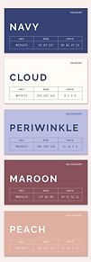
After lots of troubleshooting, I found this colour theme on Pinterest and decided to use it for my piece! I went with a different approach where the two pages have a slight gradient connection, however the second page colour is completely different.
I went through a series of MORE drafts:


Getting there...the blue colour was slightly dark so I tried lighter shades:


I finally settled on these colours where the second page was much lighter!

I changed the text organization in this section to reduce the working memory load for the reader and create a better association between the steps and small molecules.

Structure References
https://www.uniprot.org/uniprotkb/P34982/entry#structure https://alphafold.ebi.ac.uk/entry/P34982
-
Used to obtain the structure of OR1D2
https://pubchem.ncbi.nlm.nih.gov/compound/Bourgeonal
-
Used to obtain structure of bourgeonal
Other receptors/channels in the illustration:
Adenylate cyclase III: https://www.rcsb.org/structure/6YII
cAMP/cGMP CNG channel: https://www.rcsb.org/structure/7RH9
Guanylyl cyclase C: https://alphafold.ebi.ac.uk/entry/P25092